Application #22: Temporal Bone Copy
Temporal Bone
 Anatomy:
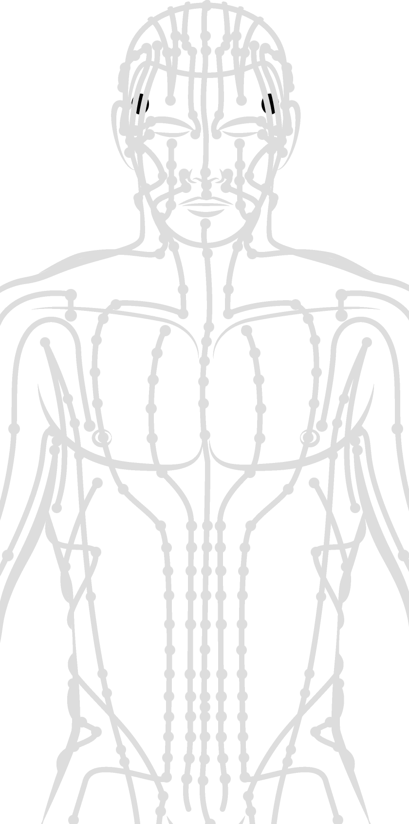
The temporal bone contributes to the lower lateral walls of the skull. It contains the middle and inner portions of the ear, and is crossed by the majority of the cranial nerves. The lower portion of the bone articulates with the mandible, forming the temporomandibular joint of the jaw.
Innervation:
The temporal bone contributes to the lower lateral walls of the skull. It contains the middle and inner portions of the ear, and is crossed by the majority of the cranial nerves. The lower portion of the bone articulates with the mandible, forming the temporomandibular joint of the jaw.
Vasculature:
The superficial temporal artery (STA) is the major branch of the temporal artery, which originates from the external carotid artery, arising above the takeoff of the internal maxillary artery. The STA then runs through the posterior portion of the parotid gland, beneath the facial nerve, and then crosses the posterior aspect of the zygomatic arch before branching into frontal and parietal branches, running within the temporalis muscle of the skull. The STA may dilate for unknown reasons and cause migraine headaches, which can be lessened with botox injections. The STA may also be involved with giant cell arteritis, an idiopathic inflammation of the artery, which necessitates a manually guided biopsy of the STA for diagnosis. This latter pathology can respond to oral steroid administration.
Possible Effects of a Strike:
- Localized pain due to location of the nervus zygomaticus, nervus auriculotemporalis, and the musculus temporalis
- damage to the arteria meningea media (middle meningeal artery)
- bleeding in the skull
- skull fracture
- traumatic brain injury